

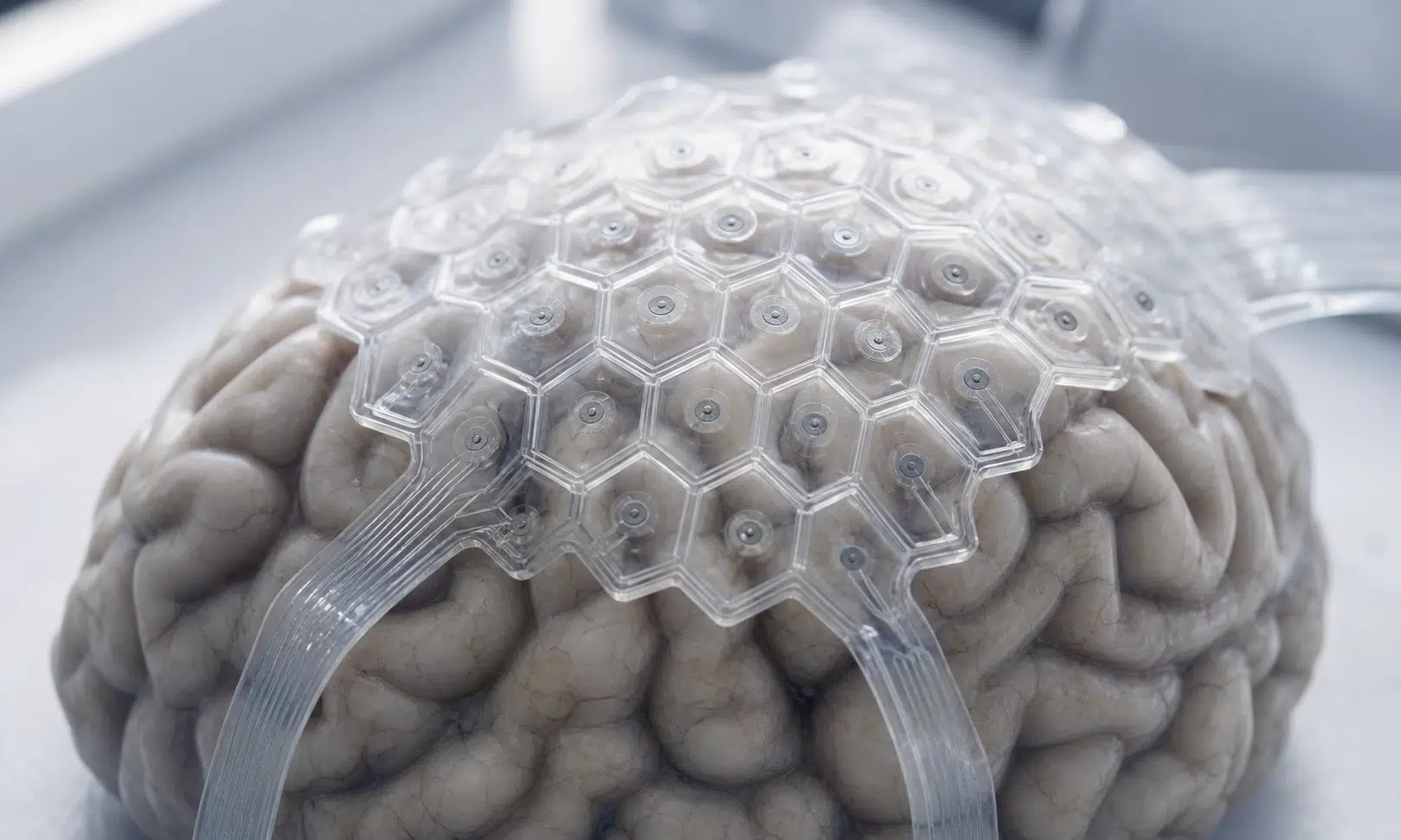

Soft 3D-printed brain sensors may offer a closer fit to the brain’s folded surface than conventional rigid devices, according to early research that combines brain mapping, flexible materials, and custom fabrication.
The work focuses on a persistent problem in surface-brain recording: no two brains fold in exactly the same way. A device that rests neatly on one person’s cortex may leave gaps on another, especially where ridges and grooves curve away from a flat electrode sheet.
That matters because surface electrodes rely on direct contact. When contact is poor, electrical signals can become weaker or noisier, making it harder to interpret brain activity clearly.
Why Brain Shape Matters
The brain’s outer surface is full of ridges and grooves. These folds help pack a large amount of neural tissue into the skull, but they also make the surface difficult to cover with a device built in a standard shape.
Researchers working with reconstructed brain scans found that fold patterns varied from one model to the next. Their results suggest that a sensor designed around one brain surface may not sit with the same precision on another.
That challenge is especially important for electrocorticography, a method that records electrical activity from the brain’s surface. Doctors use this type of recording when scalp sensors cannot provide enough detail, including in some epilepsy and movement-disorder evaluations.
In these settings, electrode contacts rest directly on the brain’s outer layer. The goal is to capture coordinated nerve-cell activity with as little interference as possible. If a pad lifts away from tissue, the recorded signal can lose clarity.
A Softer Sensor Design
The new approach uses a flexible hydrogel-based structure rather than a stiff, flat layout. Hydrogel is a water-rich material that can bend more like living tissue, which may help a device settle against curved surfaces without relying on pressure alone.
The sensor also uses an open honeycomb pattern. That structure reduces bulk while helping the device keep its shape. In practical terms, the design is meant to stay flexible without becoming too fragile to handle or use.
The research team described the honeycomb structure as a way to reduce electrode stiffness while preserving mechanical strength. That balance is important because the brain is soft, and even small pressure changes can affect tissue and recording quality.
A closer-fitting surface device could be useful in future neurological care, but the current work is still early. The findings point to a technical path, not a finished clinical product.
How Custom Printing Could Work
The process begins with a map of the brain surface. Software then uses that shape to guide the sensor pattern, so the printed device can better match a specific set of folds.
The team used direct ink writing, a type of 3D printing that deposits ink-like materials in controlled paths. The device described in the study included stacked soft layers, with insulation supporting the structure and a conductive gel layer carrying signals from tissue to recording equipment.
Because printing can produce custom shapes without many of the steps used in conventional microfabrication, the method could eventually make personalized devices more practical. That possibility is one reason the work is attracting attention: each patient’s brain surface could, in theory, guide the electrode layout.
Still, personalization adds its own hurdles. A custom device must be made reliably, cleaned or sterilized appropriately, tested for durability, and removed safely if needed. Regulators would likely examine whether a process can produce consistent quality even when each device has a different shape.
What Early Testing Suggests
In computer modeling and printed brain-surface tests, the soft honeycomb sensors appeared to sit closer to matching folds than stiffer comparison designs. The study reported smaller average gaps for the honeycomb layout, although those measurements should be understood as part of the authors’ experimental results rather than independent clinical proof.
Electrical testing also examined whether the softer design weakened signal transfer. The conductive hydrogel layer was reported to maintain low electrical resistance at the tissue-electrode interface across tested frequencies, while also supporting charge storage and delivery.
Those details matter because a sensor that fits well but cannot carry signals clearly would have limited value. The early results suggest the material approach may preserve useful electrical performance while improving mechanical fit.
Animal testing added another layer of evidence. In awake rats, the printed device recorded brain responses to brief light flashes, and the researchers compared those recordings with signals from standard electrode sets. The study reported stronger signal-to-noise performance at edge recording sites, where curved tissue often makes contact harder to maintain.
The middle recording sites showed less improvement, which fits the broader idea that flexibility may matter most where a device would otherwise lift away from the brain surface.
Safety Questions Remain
The study also looked for signs of tissue reaction after several weeks. The researchers reported no major image distortion around the device, no obvious obstruction of nearby brain-fluid movement, and no collagen scar buildup in examined tissue slices.
Those findings are encouraging, but they do not establish long-term safety in people. Rat studies, simulations, and printed models cannot answer how a human implant would behave over years of use.
Human neurosurgical devices face a much higher bar. Future work would need to address surgical handling, durability, sterilization, manufacturing control, infection risk, removal, and performance in larger brains with more complex clinical conditions.
The Path Toward Personalized Neurology
The promise of this work is not simply that a sensor can be printed. It is that brain shape, soft materials, and electrical performance can be connected in one workflow: scan the surface, design the sensor, print the device, and test whether it records more clearly.
If that workflow can be strengthened, it could support future tools for diagnosis, brain mapping, or therapies that depend on precise electrical contact. For now, the study is best read as an early step toward personalized neurology, with the most important questions still ahead.